FOLLICULAR MONITORING
It is a well-known fact that diagnostic ultrasound is a very integral part of the investigation and diagnosis of infertility. Follicular monitoring is one of the many compulsory procedures in the management of infertility. It is one of the chief components of In-Vitro Fertilization (IVF) treatment.
The Basics of IVF
IVF stands for In-Vitro Fertilization and is one of the most popular assisted reproductive techniques adopted by parents who are not able to conceive naturally due to many reasons, like advanced maternal age or damaged, or blocked, fallopian tubes.
It functions using a combination of medicines and surgical processes to fertilize egg and sperm artificially and then the fertilized egg is implanted in the mother’s uterus. The entire process of IVF takes several months to be completed. Sometimes, it works on the first try but most people have to take more than one round to see the desirable results.
IVF increases the probability of pregnancy if you are facing infertility issues but there is no guarantee of how much time would be required for it to be successful. As individuals, all of our bodies are different and hence, react differently to IVF.
Follicular Monitoring
Follicular Monitoring: What is it?
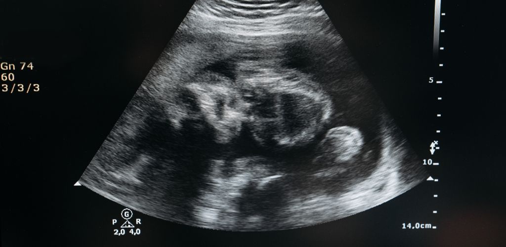
Follicular monitoring is the process of serial ultrasonic monitoring of ovarian follicles used to identify the maturity status of the eggs. It is very useful for assessing the size of the follicle that supports the growing egg and for determining the thickness of the uterine lining. It also helps in documenting the ovulation path.
A follicle is an aggregation of cells that are found in a woman’s ovaries. Each follicle releases 1 egg in a lifetime. Women are born with roughly 400,000 follicles. Follicular study and pregnancy are closely related. A follicular study is carried out to get a complete picture of whether a woman is ovulating at the time.
Follicular tracking involves the study of the development of the follicle, to find out how close to ovulation a woman is. It includes a series of vaginal scans that help to identify the stage of the woman’s current menstrual cycle.
Why is it done?
- Follicular monitory is very important in most fertility This to track how a woman’s body reacts to those treatments.
- If a couple opts for IVF, it is important to conduct follicular scans. This is to assess the health and number of eggs produced in each ovulation.
- Follicular scanning also helps us to keep a track of the level of concerning hormones.
- In the case of decreased levels of hormones, we can adjust the dose accordingly in a way that increases the chances of conception.
- Hyperresponsiveness can also be taken care of, as observed in some patients.
- Using these scans, we can time Intrauterine Insemination and avoid Ovarian Hyperstimulation Syndrome and multiple pregnancies by regulating doses of hormones.
How is it done?
- By ultrasound scanning, colour doppler, power doppler-morphological growth of follicles.
- Estradiol Alone- it indicates the functional activities of the follicles.
- Using transvaginal Ultrasound.
Follicular Monitoring Ultrasound:
- Ultrasound scanning is the method by which the follicular monitoring procedure.
- The ovarian follicles are thoroughly examined through pictures of the internal organs that are taken by inserting small plastic probes into the vagina. The process is carried out by certified sonographers.
- The woman will need to lie down in the stirrup position so that the scan can be done. With a sheet covering her from the waist down, the probes are inserted.
- These probes emit sound waves in the ultrasound frequencies, using which the images are captured.
- The sonographer will be able to approximately predict the time the egg will be released, depending upon the thickening of the walls of the uterus.
- She can, hence, plan your efforts to conceive to maximize your chances. The follicular study process is, therefore, of great use if you are trying to have a child.
Methods of Follicular Monitoring (TAS vs TVS)
Ultrasound has the unique ability to document the morphologic changes in the ovary and uterus during normal and induced cycles. Several studies were done in the early eighties regarding the sonographic evaluation of normal and induced ovulation.
With the introduction of transvaginal ultrasonography, between 1983-1985 the scenario started to change rapidly. Transvaginal sonography (TVS) started to gradually gain acceptance among radiologists and obstetricians. Several investigators showed that transvaginal scanning improved the visualisation of follicular structure in comparison with transabdominal (TA) scanning in the majority of patients.
Transabdominal ultrasound was initially utilized in its most colloquial setting of pregnancy in the 1960s. It is now utilized for visualization of multiple abdominal organs. Transabdominal sonography (TAS) images the pelvic organs through the anterior abdominal wall in the supra-pubic region.
There are two major limitations of TAS. Firstly, there is a need to use lower frequencies for imaging because of the long distance between the transducer and the pelvic organs. The other disadvantage is the presence of a beam degrading effect of the anterior abdominal wall, especially in obese patients. Both these limitations result in a fall in the image quality.
On the other hand, Transvaginal Sonography (TVS) is a procedure used to examine the vagina, uterus, fallopian tubes, ovaries, and bladder.
An instrument is inserted into the vagina which causes sound waves to bounce off organs inside the pelvis. These sound waves create echoes that are sent to a computer that creates a picture called a sonogram.
Transvaginal sonography (TVS) produces greatly improved resolution as compared to TAS, primarily due to the higher frequencies employed and also due to the absence of beam deformation by the anterior abdominal wall. Major advantages of TVS over TAS are - improved image quality and avoidance of patient discomfort due to full urinary bladder.
Frequently Asked Questions:
- Who needs to get the follicular monitoring done?
It is advisable to get the follicular monitoring done for the following cases:
- Couples who are trying to get pregnant but have not succeeded even after trying for many months.
- Women who are unsure of their ovulation despite using prediction kits.
- Women who have experienced unfortunate pregnancies in the early phases of pregnancies. This can help them understand the reason for miscarriages so that they can take care of future pregnancies.
- Women who have been taking drugs or medicines for any conception-based issues.
- How long does the follicular tracking/monitoring scan take?
The process of scanning itself takes less than a quarter of an hour but preparation for the scan may take up a couple of hours before the scanning takes place. If coordination between the patient and doctor and further with the sonographer is smooth, the scanning procedure is not more than fifteen minutes long.
- How many scans are carried out in a single cycle?
It is observed that it is common to carry out four to six scans during a cycle, to better ascertain when ovulation will take place. The initial scan is known as the baseline scan, and it helps the doctor understand the initial stage of the follicle with great certainty. Further, the doctor schedules the scans at the optimal times to follow the development of the uterine follicles. This is done to maximize the chances of ovulation and hence catalyzes the procedure of conception.
- What else can be detected using a follicular ultrasound scan?
Even though this is mainly used for tracking the growth of eggs, follicular scanning can be used for figuring out the root cause of not being able to conceive.
- The doctor will be able to identify the follicles using this scan that does not grow properly and rupture at an early stage.
- The scan also finds out if the follicles are not growing at all or are not rupturing at the right time to allow the fertilization of the egg.
- How can we predict the success rate of IVF using scanning?
Most of the IVF studies are conducted after the induction of ovaries using ovulation using agents like Clomiphene Citrate. The main success-determining factors are:
- Ovarian Volume
- Antral Follicle Number
- Ovarian Stromal Blood Flow
- Can there be any side effects of follicular scanning?
- There are no physical side effects that can occur to you as a result of this scan, but it can be a taxing and exhausting ordeal for most couples.
- Most couples who are prescribed to this process would have been trying hard to conceive a baby for a few months now, so these scans may cause sexual compatibility problems between them – which can lead to marital disharmony.
- Since doctors, on basis of previous scans, would tell the exact time of ovulation, the woman may want to have sex only during that particular period of ovulation. This can create a sexual drift and also affect male partner psychologically. He may feel more of a sperm donor and less of a sexual/romantic partner.
- The regular scans, taking leaves and missing meetings, and mental stress can lead to not just personal conflicts but also career pressure.
There can be many reasons behind not being able to conceive and it is important to track those root causes. The follicular ultrasound scanning enables you to figure out the reasons and do your best to avoid those in future pregnancies. It can be difficult to deal with the stress but the satisfaction of having your baby is unexplainable.
References:
https://www.ncbi.nlm.nih.gov/pmc/articles/PMC5531945/
https://www.ncbi.nlm.nih.gov/pmc/articles/PMC4838583/
https://www.ncbi.nlm.nih.gov/books/NBK534813/
https://www.ncbi.nlm.nih.gov/pmc/articles/PMC5532046/
By -
Dr. Ruchika Singh
14-April-2023